File:1562466022013-lg.jpg
Jump to navigation
Jump to search

Size of this preview: 800 × 531 pixels. Other resolutions: 320 × 212 pixels | 2,008 × 1,332 pixels.
Original file (2,008 × 1,332 pixels, file size: 968 KB, MIME type: image/jpeg)
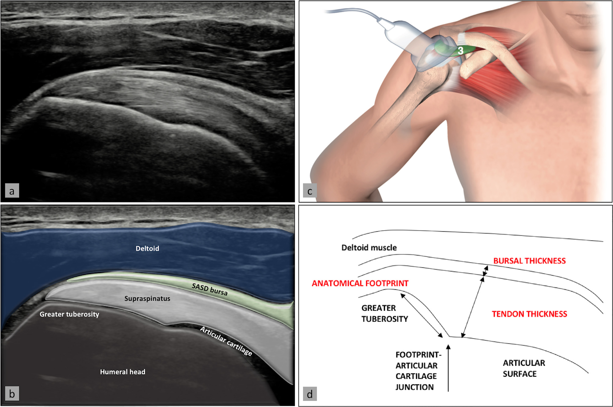
Supraspinatus tendon, visualized on its longitudinal axis, and the subacromial-subdeltoid bursa lying superficially to it. Ultrasound image (a) with superimposed anatomy (b), patient/probe position (c), and landmarks for measurement of these two structures (d). Reproduced from Plomb-Holmes et al., with permission
File history
Click on a date/time to view the file as it appeared at that time.
| Date/Time | Thumbnail | Dimensions | User | Comment | |
|---|---|---|---|---|---|
| current | 16:47, 2 January 2020 | | 2,008 × 1,332 (968 KB) | Alexandre.laedermann (talk | contribs) |
- You cannot overwrite this file.
File usage
The following 3 pages uses this file: