File:1563010262654-lg.jpg
Revision as of 16:17, 7 January 2020 by Alexandre.laedermann (talk | contribs)

Size of this preview: 800 × 411 pixels. Other resolutions: 320 × 164 pixels | 2,982 × 1,532 pixels.
Original file (2,982 × 1,532 pixels, file size: 2.79 MB, MIME type: image/jpeg)
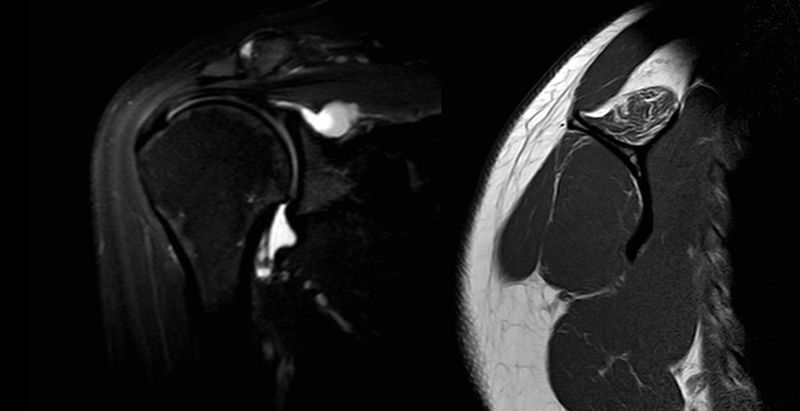
Left: MRA coronal view with T2 sequences revealing a large spinoglenoid cyst. Right: MRI sagittal view of the same patient with T1 sequences of a fatty infiltrated supraspinatus.
File history
Click on a date/time to view the file as it appeared at that time.
| Date/Time | Thumbnail | Dimensions | User | Comment | |
|---|---|---|---|---|---|
| current | 16:17, 7 January 2020 | | 2,982 × 1,532 (2.79 MB) | Alexandre.laedermann (talk | contribs) |
- You cannot overwrite this file.
File usage
The following 2 pages uses this file: