File:Figure 15 jpeg.jpg
Jump to navigation
Jump to search
Size of this preview: 800 × 288 pixels. Other resolutions: 320 × 115 pixels | 3,360 × 1,208 pixels.
Original file (3,360 × 1,208 pixels, file size: 578 KB, MIME type: image/jpeg)
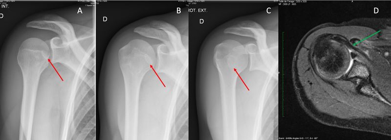
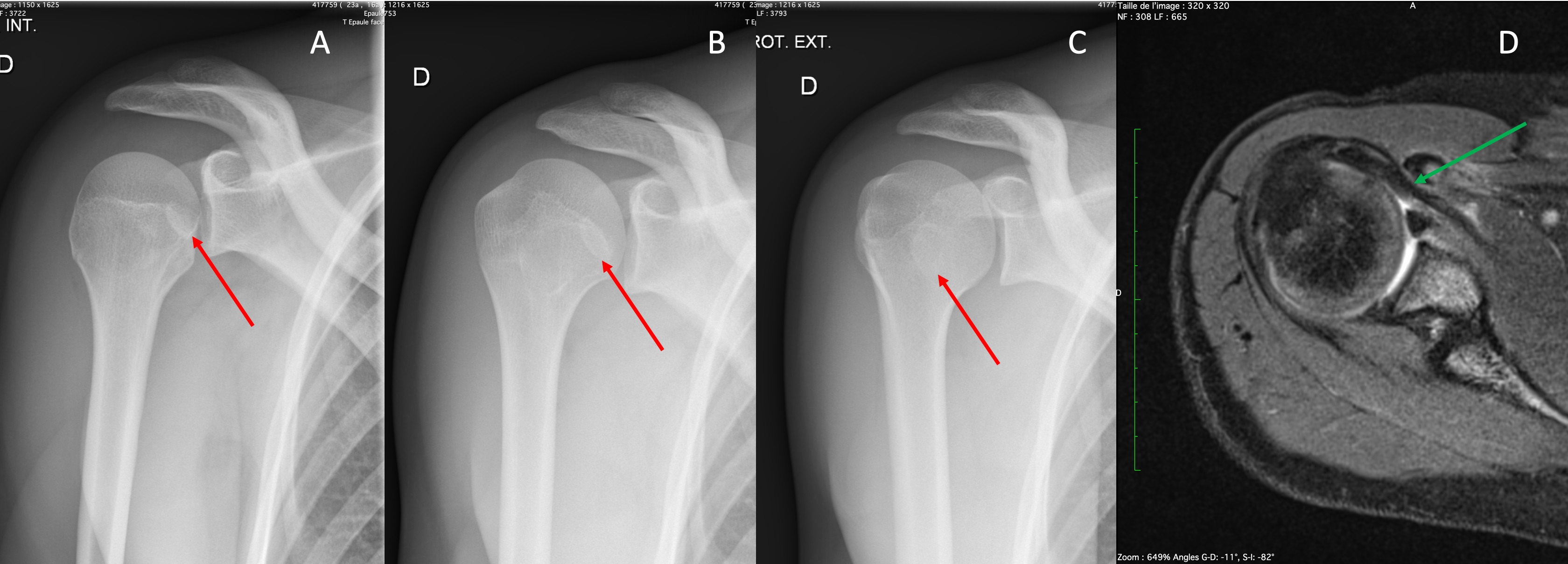
X-ray in internal (A), neutral (B) and external rotation (C) of a 14-year-old patient that sustain a trauma 2 years before demonstrating pseudarthrosis of the coracoid process with a bony fragment adherent to the subscapularis (red arrows). Axial T2 fat saturation sequence magnetic resonance imaging (MRI) of the same patient showing a narrowing of the subcoracoid space.
File history
Click on a date/time to view the file as it appeared at that time.
| Date/Time | Thumbnail | Dimensions | User | Comment | |
|---|---|---|---|---|---|
| current | 11:03, 2 June 2021 | 3,360 × 1,208 (578 KB) | Alexandre.laedermann (talk | contribs) |
- You cannot overwrite this file.
File usage
The following page uses this file: