File:Classification lésion labrale nourissat.png

Original file (2,166 × 1,686 pixels, file size: 1.35 MB, MIME type: image/png)
- Example.jpg
Caption1
- Example.jpg
Caption2
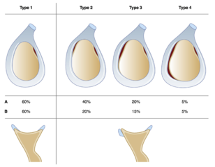
Type 1: the most frequent, corresponding to the direct insertion of the posterior labrum in the cartilage surface, without a gap. It represents 60% of shoulders. In type 2 there is no direct contact between cartilage surface and the superior segment of the posterior labrum, with a medialized aspect of the segment. This aspect is frequent (40%) and isolated in 20% of cases. It can be associated with medialization of the medial fragment in 15% of cases corresponding to type 3. Type 4: (5%) all labrum is medialized. A: percentage of medialized aspect of the posterior labrum, by segment. B: percentage of each type of insertion. Bottom: the two modalities of fixation of the posterior labrum: left, in continuity with articular surface; right, medialized aspect, with a gap between the labrum and the cartilage. Reproduced from Nourissat et al.,with permission.
File history
Click on a date/time to view the file as it appeared at that time.
| Date/Time | Thumbnail | Dimensions | User | Comment | |
|---|---|---|---|---|---|
| current | 08:41, 31 December 2019 | | 2,166 × 1,686 (1.35 MB) | Alexandre.laedermann (talk | contribs) |
- You cannot overwrite this file.
File usage
The following file is a duplicate of this file (more details):
There are no pages that use this file.