File:1562914895053-lg.jpg
Jump to navigation
Jump to search
Size of this preview: 800 × 301 pixels. Other resolutions: 320 × 120 pixels | 2,848 × 1,072 pixels.
Original file (2,848 × 1,072 pixels, file size: 2.23 MB, MIME type: image/jpeg)
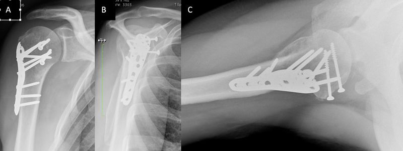
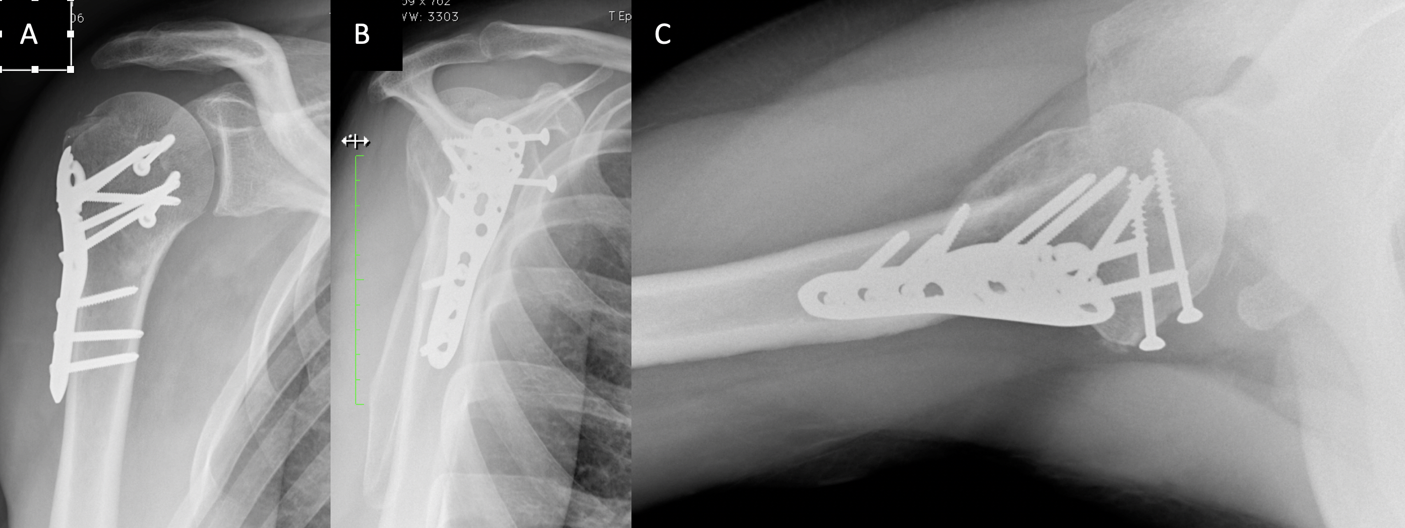
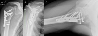
Figure 19. Frontal (A), Lamy lateral (B) and axial (C) radiographs, 6 weeks after a type II right-sided cephalotubercular ‘head split’ fracture. Note the two anteroposterior malleolar screws that permit compression of the head fracture, while the plate stabilizes the lesser tuberosity fracture.
File history
Click on a date/time to view the file as it appeared at that time.
| Date/Time | Thumbnail | Dimensions | User | Comment | |
|---|---|---|---|---|---|
| current | 20:30, 7 January 2020 | 2,848 × 1,072 (2.23 MB) | Alexandre.laedermann (talk | contribs) |
- You cannot overwrite this file.
File usage
The following 3 pages uses this file: