File:1562483248519-lg.jpg
Jump to navigation
Jump to search

Size of this preview: 690 × 600 pixels. Other resolutions: 276 × 240 pixels | 1,058 × 920 pixels.
Original file (1,058 × 920 pixels, file size: 358 KB, MIME type: image/jpeg)
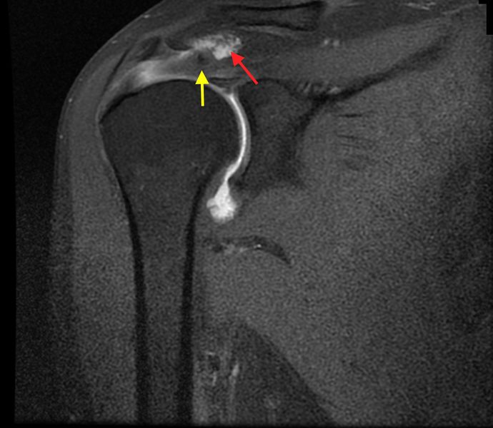
Coronal T1 weighted MRA image with fat saturation of a right shoulder demonstrates adhesions between the bursal side of the tendon and the wall of the subacromial bursa (red arrow), and abnormal orientation of the fibers stump (yellow arrow). Reproduced from Lädermann et al., with permission.
File history
Click on a date/time to view the file as it appeared at that time.
| Date/Time | Thumbnail | Dimensions | User | Comment | |
|---|---|---|---|---|---|
| current | 12:24, 3 January 2020 | | 1,058 × 920 (358 KB) | Alexandre.laedermann (talk | contribs) |
- You cannot overwrite this file.
File usage
The following 3 pages uses this file: