File:1562645724133-lg.jpg
Revision as of 20:08, 30 December 2019 by Alexandre.laedermann (talk | contribs)

Size of this preview: 483 × 599 pixels. Other resolutions: 193 × 240 pixels | 950 × 1,178 pixels.
Original file (950 × 1,178 pixels, file size: 342 KB, MIME type: image/jpeg)
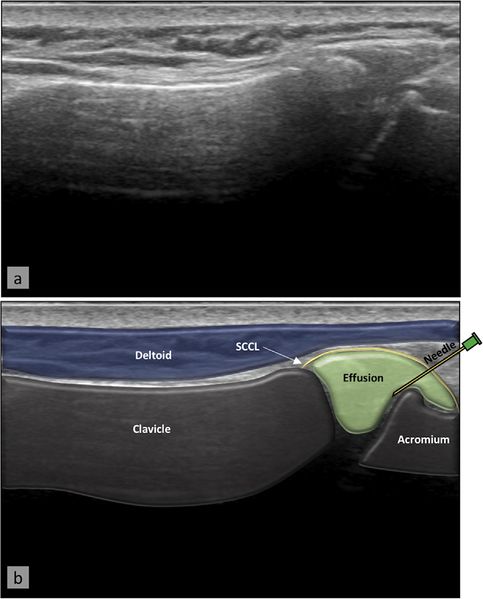
Acromioclavicular joint injection. The needle is inserted at the mid-line of the lateral edge and parallel to the probe, allowing it to be visualized entering the acromioclavicular joint. US image (a) with superimposed anatomy (b). From Plomb-Holmes et al., with permission.
File history
Click on a date/time to view the file as it appeared at that time.
| Date/Time | Thumbnail | Dimensions | User | Comment | |
|---|---|---|---|---|---|
| current | 20:08, 30 December 2019 | | 950 × 1,178 (342 KB) | Alexandre.laedermann (talk | contribs) |
- You cannot overwrite this file.
File usage
The following page uses this file: