File:1562643120067-lg.jpg

Original file (943 × 429 pixels, file size: 366 KB, MIME type: image/jpeg)
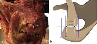
Figure. 1 Photograph (a) and drawing (b) showing measurement methods. The anterior (A) and posterior (P) borders of the capsule are marked and connected with a straight line (AP line). Two parallel lines perpendicular to the AP line are created that passed through points A and P. Two anterior parallel lines perpendicular to the AP line are drawn: one passing at the most anterior edge of the acromion (acromion anteriorly [ACA]) and the other passing at the most anterior edge of the clavicle (clavicle anteriorly [CLA]). The same procedure is followed for the posterior part of the joint, and the lines passing from the acromion posteriorly (ACP) and clavicle posteriorly (CLP) were drawn. Reproduced with permission from Barth et al.
File history
Click on a date/time to view the file as it appeared at that time.
| Date/Time | Thumbnail | Dimensions | User | Comment | |
|---|---|---|---|---|---|
| current | 14:02, 24 December 2019 | | 943 × 429 (366 KB) | Simon.de chabaneix (talk | contribs) |
- You cannot overwrite this file.
File usage
The following 3 pages uses this file: