File:1562484142491-lg.jpg
Revision as of 12:53, 3 January 2020 by Alexandre.laedermann (talk | contribs)

Size of this preview: 800 × 381 pixels. Other resolutions: 320 × 153 pixels | 875 × 417 pixels.
Original file (875 × 417 pixels, file size: 194 KB, MIME type: image/jpeg)
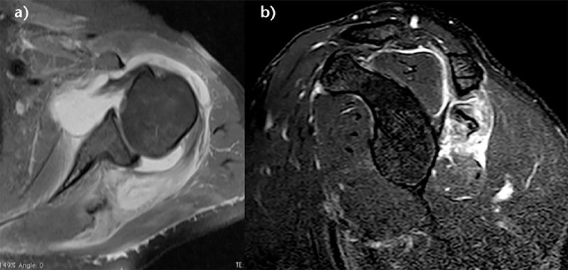
Axial and sagittal T2-weighted FATSAT magnetic resonance imaging (MRI) images demonstrating a type C rotator cuff lesion with an intact tendon, a stage 3 rupture of the musculotendinous junction, and huge edema of the muscle. Reproduced from Lädermann et al., with permission.
File history
Click on a date/time to view the file as it appeared at that time.
| Date/Time | Thumbnail | Dimensions | User | Comment | |
|---|---|---|---|---|---|
| current | 12:53, 3 January 2020 | | 875 × 417 (194 KB) | Alexandre.laedermann (talk | contribs) |
- You cannot overwrite this file.
File usage
The following 3 pages uses this file: