File:Figure 7 jpeg.jpg
Revision as of 11:08, 2 June 2021 by Alexandre.laedermann (talk | contribs)
Size of this preview: 600 × 600 pixels. Other resolutions: 240 × 240 pixels | 1,824 × 1,824 pixels.
Original file (1,824 × 1,824 pixels, file size: 329 KB, MIME type: image/jpeg)
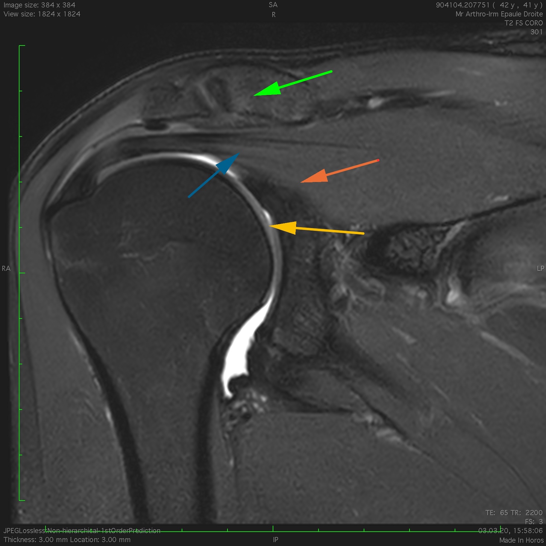
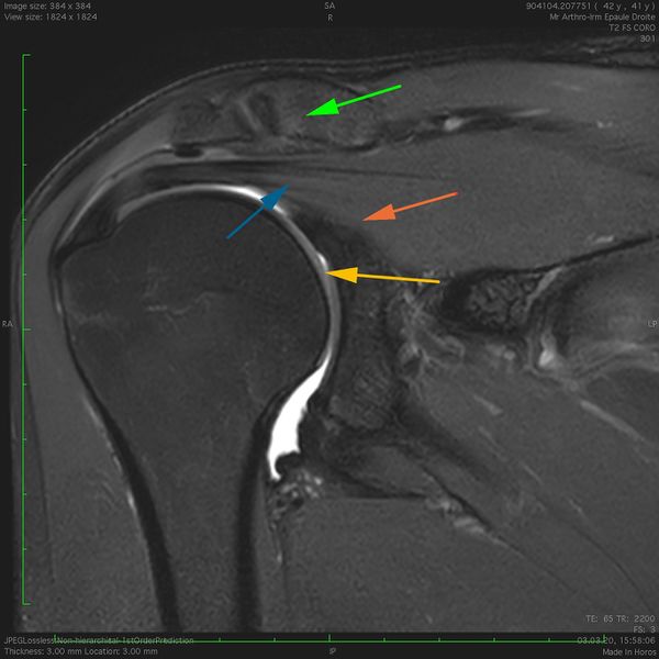
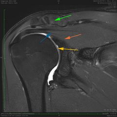
Sagittal magnetic resonance imaging (MRI) T2 fat saturation view of a right subcoracoid space, the coracoid process (green arrow) defines the superior border, the conjoint tendon (yellow arrow) form the anterior border, the glenoid (blue arrow) and the middle glenohumeral ligament (red arrow) serve as the posterior border.
File history
Click on a date/time to view the file as it appeared at that time.
| Date/Time | Thumbnail | Dimensions | User | Comment | |
|---|---|---|---|---|---|
| current | 11:08, 2 June 2021 | | 1,824 × 1,824 (329 KB) | Alexandre.laedermann (talk | contribs) |
- You cannot overwrite this file.
File usage
The following page uses this file: