File:Figure 5 jpeg.jpg
Revision as of 10:51, 2 June 2021 by Alexandre.laedermann (talk | contribs)
Size of this preview: 600 × 600 pixels. Other resolutions: 240 × 240 pixels | 1,824 × 1,824 pixels.
Original file (1,824 × 1,824 pixels, file size: 288 KB, MIME type: image/jpeg)
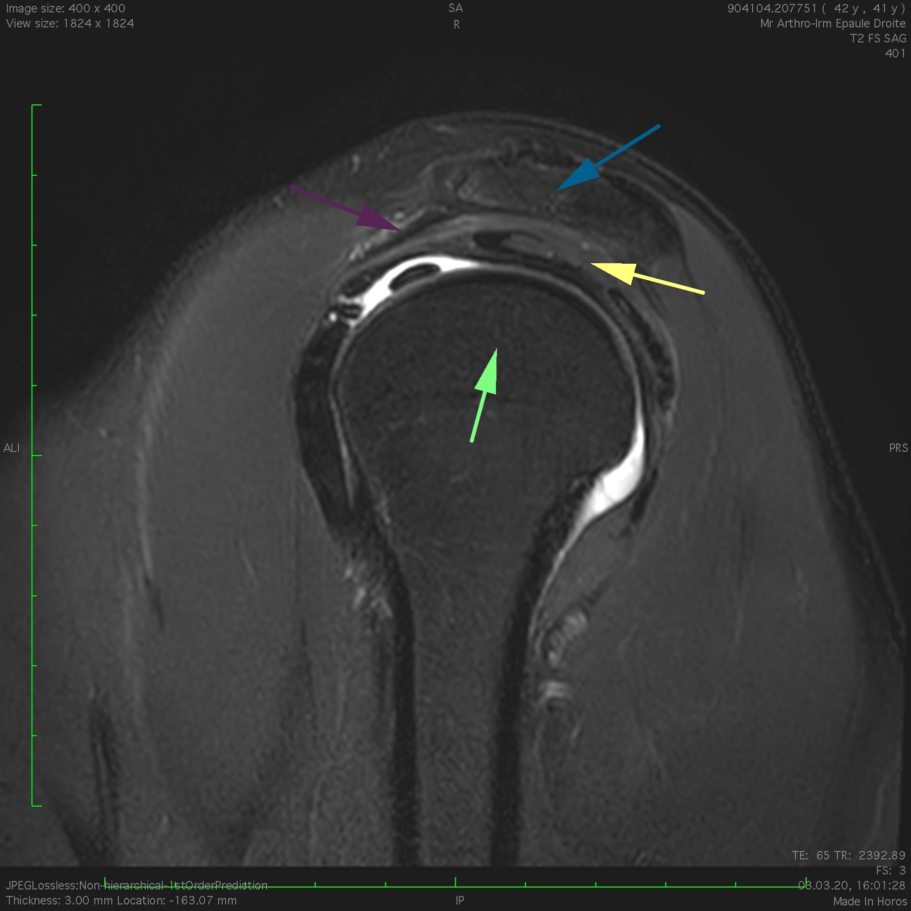
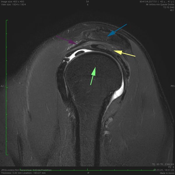
Magnetic resonance imaging (MRI) T2 fat saturation sagittal view of a right subacromial space. The acromion superiorly (blue arrow), the coracoacromial ligament anteriorly (purple arrow) and the humeral head inferiorly (green arrow) surround the supraspinatus tendon (yellow arrow). Note: anterior is the left in the image.
File history
Click on a date/time to view the file as it appeared at that time.
| Date/Time | Thumbnail | Dimensions | User | Comment | |
|---|---|---|---|---|---|
| current | 10:51, 2 June 2021 | | 1,824 × 1,824 (288 KB) | Alexandre.laedermann (talk | contribs) |
- You cannot overwrite this file.
File usage
The following page uses this file: