File:1562442208871-lg.jpg

Original file (1,744 × 705 pixels, file size: 216 KB, MIME type: image/jpeg)
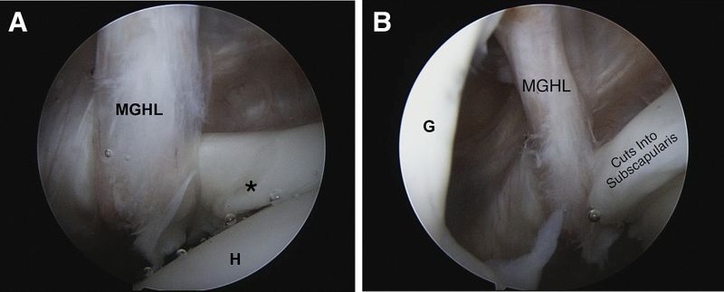
(A) Posterior portal view of the right shoulder with a 30_ arthroscope. The humeral head (H) is on the lower right. The MGHL (labeled) sits approximately 1 cm medial to a significant abrasion lesion of the upper boarder of the subscapularis (*). (B) Posterior portal view of the right shoulder with a 30_ arthroscope. The glenoid (G) is visible on the left side of the field of view. With internal rotation of the humerus, the MGHL (labeled) can clearly be seen to be the offending lesion cutting into the upper subscapularis causing the pathology in the subscapularis. (MGHL, middle glenohumeral ligament.) Reproduce from Brady et al., with permission.
File history
Click on a date/time to view the file as it appeared at that time.
| Date/Time | Thumbnail | Dimensions | User | Comment | |
|---|---|---|---|---|---|
| current | 16:40, 3 January 2020 | 1,744 × 705 (216 KB) | Alexandre.laedermann (talk | contribs) |
- You cannot overwrite this file.